La recherche
Depuis sa création, l’Institut Jules Bordet n’a cessé de chercher à comprendre les mécanismes cellulaires impliqués dans le cancer et à améliorer les méthodes diagnostiques et les traitements anticancéreux. En collaborant avec des chercheurs et des médecins de tous horizons, les chercheurs de l’Institut Jules Bordet transfèrent au chevet des patients les innovations issues de leurs recherches scientifiques.

« Les projets menés par les chercheurs de l’Institut Jules Bordet se distinguent par leur transdisciplinarité et par leur inventivité. Faire progresser le soin et explorer toutes les pistes de recherche possibles : telle est la démarche de l’Institut Jules Bordet. »
Titre
Comment la recherche fait avancer le traitement des cancers
A l’Institut Jules Bordet, la recherche est à la fois clinique et translationnelle. Cette activité très organisée mobilise et engage de nombreux professionnels de la santé ainsi que les patients.
La Recherche Clinique
La recherche clinique, ce sont les études cliniques que nous menons à l’Institut Jules Bordet avec nos partenaires, centres de recherche académiques et entreprises pharmaceutiques. Les objectifs de ces études cliniques visent, par exemple, à comparer plusieurs stratégies thérapeutiques ou à tester de nouvelles molécules dans des cancers où il n’existe pas de traitement suffisamment efficace à ce jour. La recherche clinique n’est possible qu’avec l’engagement des patients. Leur participation aux essais cliniques permet aux chercheurs de répondre à d’importantes questions de recherche qui serviront ensuite d’autres patients.
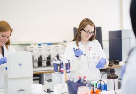
La Recherche Translationnelle
Comprendre le cancer, rechercher des indicateurs ou des marqueurs qui aideront au diagnostic et au traitement ou encore à l’évaluation du pronostic du patients sont les objectifs principaux de la recherche dite translationnelle. Ce type de recherches se fait le plus souvent sur des échantillons prélevés lors du diagnostic ou du traitement des patients, avec leur accord. Intégrée à la recherche clinique, la recherche translationnelle a pour ambition de concrétiser rapidement les découvertes faites au laboratoire dans les soins proposés aux patients.
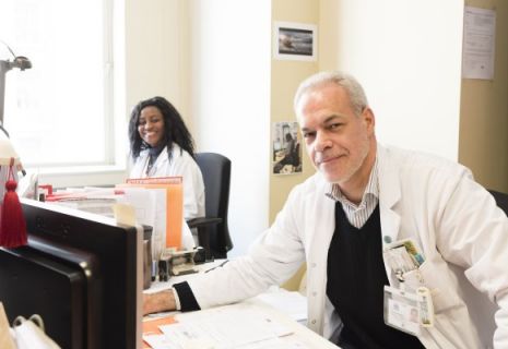
Conduire et organiser la recherche clinique
L’Institut Jules Bordet participe à la recherche contre le cancer en menant des essais cliniques conçus par ses chercheurs ou d’autres groupes de recherche à travers le monde. Plusieurs structures participent au sein de l’Institut à l’organisation et à la conduite des essais cliniques :
- Le Comité d’Ethique : un comité indépendant qui analyse et évalue les projets de recherche avant qu’ils ne soient proposés aux patients afin de s’assurer qu’ils respectent les règles éthiques.
- Le Support operationnel promotion (CTC – CTSU) : une unité de soutien qui organise les différents aspects, en particulier règlementaires (compliance aux bonnes pratiques cliniques, pharmacovigilance…), et assure la qualité des études cliniques initiées par l’Institut avec les partenaires de la recherche.
- La Clinical Trials Conduct Unit (CTCU) : une unité composée d’infirmiers de recherche, de data managers et de médecins-investigateurs qui suivent les patients participant à des essais cliniques au sein de l’Institut. Cette unité travaille en étroite collaboration avec tous les services de l’Institut dont l’oncologie médicale, l’hématologie, la psycho-oncologie, etc…
- Le Support contractuel promotion et investigation (CTC) : une unité centralisée de gestion administrative des projets de recherche qui aide les chercheurs d’un point de vue juridique, contractuel, suivi financier….
Collaborer pour innover
La recherche est toujours une activité collaborative, particulièrement dans le domaine du cancer. Pour explorer les pistes les plus prometteuses, l’Institut Jules Bordet s’associe avec d’autres partenaires académiques et/ou des entreprises privées tant en Belgique qu’à l’étranger.
Partenaires académiques
La recherche académique est inscrite dans l’ADN de l’Institut Jules Bordet. Nous sommes d’ailleurs à l’origine de prestigieux réseaux de recherche internationaux qui ont vu le jour à l’Institut : la European Organization for Research and Treatment of cancer (EORTC), le Breast International Group (BIG) et plus récemment le réseau Oncodistinct. Proposer les meilleurs soins pour chaque patient est une entreprise délicate, compte tenu de la complexité et de la diversité des situations cliniques et des stratégies thérapeutiques possibles. Les soignants doivent donc interroger leur pratique de façon rigoureuse, notamment au moyen d’essais cliniques. Ceux-ci s’organisent dans un cadre universitaire, en particulier avec l’Université Libre de Bruxelles (ULB) et la VUB. Leurs résultats ont souvent un impact important dans les soins proposés aux patients.
Partenaires privés
Pour trouver un nouveau médicament, une nouvelle méthode diagnostique ou un nouveau dispositif médical pour ses patients, l’Institut Jules Bordet soutient et collabore avec ceux qui développent ces techniques et traitements innovants : les entreprises pharmaceutiques, de biotechnologies ou de dispositifs médicaux mais aussi des start up. Certains essais cliniques menés à l’Institut Jules Bordet se font donc en partenariat avec ces entreprises et toujours dans le respect et l’intérêt du patient.
Titre
Participer aux découvertes contre le cancer
Les essais cliniques
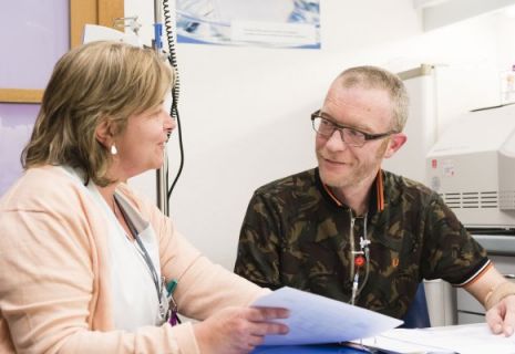
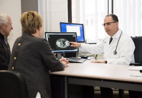
En tant que patient(e), vous serez peut-être invité(e) à participer à un essai clinique portant sur un nouveau traitement, une nouvelle technique diagnostique ou une approche innovante en terme de soins. Outre de possibles bénéfices pour vous-même, participer à un essai clinique contribue aux progrès de la médecine et aide les futurs patients.
Cependant, votre participation à ces études est strictement volontaire et non obligatoire. Aucune démarche de recherche ne sera jamais réalisée sans votre accord. Afin de pouvoir décider en toute connaissance de cause et donner ce qu’on appelle le « consentement éclairé du patient », vous recevrez une information claire et compréhensible, oralement et par écrit. Votre médecin référent vous détaillera notamment les possibles risques et bénéfices relatifs à la participation à l’essai clinique qui vous est proposé. Les infirmiers de recherche sont également là pour répondre à toutes les questions que vous vous poseriez.
Votre santé et votre qualité de vie sont au cœur de nos préoccupations. Si votre état de santé l’exige ou si c’est votre souhait, vous êtes libre d’interrompre votre participation à l’essai clinique à tout moment.