At the Anatomical Pathology Department of the Jules Bordet Institute expert pathologists are increasingly turning to digital pathology, a truly essential "e-health" tool for analysing tumours and the tumour microenvironment.
It is the digitisation of microscopic slides on which samples (operative specimens, biopsies, cytological and blood samples, etc.) are placed.
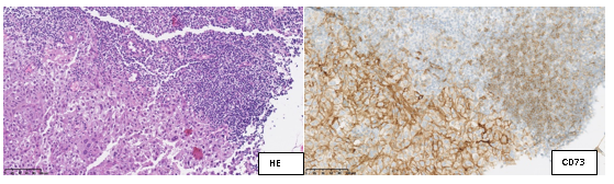
Synergy clinical study : metastatic lymph node (breast carcinoma)
These slides are digitised using the latest generation scanners, at the cutting edge of technology. It is thanks to the combined efforts of pathologists and IT experts that it has been possible to develop and use these "modern microscopes". They are able to digitise, annotate and classify the slides on platforms that are easily accessible and totally secure. Digital pathology facilitates the painstaking work of analysing cells and biological tissue that is the day-to-day work of pathologists. The Anatomical Pathology Department of the Jules Bordet Institute is committed to this development that marks a new stage in data management and computerisation.
Digital pathology has a number of benefits :
- It permits a morphological images storage that over time is more efficient and of a higher quality. The slides are digitised and no longer need to be conserved physically, thereby avoiding any loss of slide quality (over time).
- It represents the future of histopathology (the tissue analyse with a microscope) through a more precise diagnosis thanks to the various possibilities offered by artificial intelligence.
- It permits an easy and secure data sharing with other experts to arrive at a medical opinion, including across the globe.
The images digitally generated using the scanner at the Jules Bordet Institute Department of Anatomical Pathology are presented, discussed and compared to the PET-FDG, CT, MRI and other images at the Multidisciplinary Cancer Conferences, where experts come together to discuss individual cases.
In the event of diagnostic doubt, a second opinion can be requested remotely. This is known as telepathology, that is, the sharing of information with other experts to refine the diagnosis. At the Jules Bordet Institute our skills are also employed to review different clinical case studies. Thanks to the principle of cloud computing, the online sharing of virtual slides makes it possible to analyse resources originating at different and/or geographically distant laboratories. Users connect to a portal that gives them controlled and secure access to virtual slides. They can then share or annotate them, for example, directly online.
Pathologists at the Jules Bordet Institute have become accustomed, over time, to creating digital image libraries of interesting cases. The image quality is optimal and classification and access are fast and easy, dispensing with the need to retrieve glass slides from the archives (the Anatomical Pathalogy Department already has hundreds of thousands of glass slides). These rare cases also serve as a teaching and reference aid in case of doubt when reaching a diagnosis.
Digital slides are an essential tool for training young pathologists and for presentations at conferences and seminars.
In addition, many tools are integrated in the readers to increase the educational or diagnostic potential of the digital slide. These include the possibility of attaching annotations or comments. It is also possible to exceed the optical magnification limits of the microscope or scanner using an automatic zoom. A "multi-window" system also makes it possible to view several slides simultaneously: their synchronised movement is very useful for observing several different colours on the fields.
The slide scanner is used currently in our scientific activities and publications due to the excellent image resolution and the possibility of annotating the tissue components.
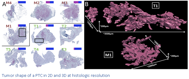
Mention can also be made of projects for the ultra-rapid visualisation of virtual slides in 3D by digitising hundreds of successive tissue sections for the purpose of exploring the thickness of the image produced. This type of analysis makes it possible to evaluate the volume of the structures analysed by the pathologist and not just their surface.
Pathologists at the Jules Bordet Institute, together with researchers and bioinformaticians, analyse digital slides on large high definition screens. This enables the pathologist to make very precise annotations, which are today essential in spatial transcriptomics. This technique makes it possible to analyse the gene expression at several hundred points within a tissue section. Specifically, these are small microarrays (6 × 6 mm2) including a thousand plots. Each plot provides information on all the genes expressed, whereas in situ hybridization only examines one gene at a time. The localisation of these spots on the slide is essential for the pathologist's results analysis.
The slide scanner is therefore a tool that, with the participation of experts, could automate activities and favour the development of Artificial Intelligence. The information made available to the pathologist is concentrated and reliable and makes the diagnosis more rapid and more precise. Many image analysis research projects are seeking to study the validity of these algorithms in regard to a wide variety of images. Our department is actively involved in two networks, one in the USA (FDA) and the other European (TIGER).
To perfect our analyses, we are equipped with a digital image processing program. Many applications are now available: TIL quantification per population, ki67 scoring, the detection of tumour zones, surface area measurements, etc. These applications can be used in research but also when completing or validating a diagnosis.
Research
Publications, congress presentations, projects...
Virtual ESMO Annual 2020 congress
First findings from SYNERGY, a phase I/II trial testing the addition of the anti-CD73 oleclumab to the anti-PD-L1 durvalumab and chemotherapy as 1st line therapy for patients with metastatic triple-negative breast cancer
D. Eiger, C. Maurer, M. Brandão, P. Aftimos, K. Punie, D.Taylor, T. Van den Mooter, R.Poncin, JL. Canon, F. Duhoux, V. Casert, F. Clatot, C.Velghe, L. Craciun, M. Paesmans, E. de Azambuja, M. Ignatiadis, D. Larsimont, M. Piccart-Gebhart, L. Buisseret
Best oral presentation-Belgian Week of Pathology 2021
Implementation of automatic quantification of Ki-67 in well- differentiated gastro-entero-pancreatic neuroendocrine tumors: towards standardized evaluation into daily practice.
F. Lifrange, N. Gumus, L. Craciun, M. Gomez Galdon, P. Demetter, L. Verset.
Breast 2020 Dec; 54:179-186
Digital analysis of distant and cancer-associated mammary adipocytes.
E. Isnaldi, F. Richard, M. De Schepper, D. Vincent, S. Leduc, M. Maetens, T. Geukens, G. Floris, G. Rouas, F. Cardoso, C. Sotiriou, G. Zoppoli, D. Larsimont, E. Biganzoli, C. Desmedt.
J Clin Endocrinol Metab 2018
Distinctive Desmoplastic 3D Morphology Associated With BRAFV600E in Papillary Thyroid Cancers.
M. Tarabichi, A. Antoniou, S. Le Pennec, D. Gacquer, N. de Saint Aubain, L. Craciun, T. Cielen, I. Laios, D. Larsimont, G. Andry, JE. Dumont, C. Maenhaut, V. Detours.
San Antonio Breast Cancer Symposium 2021
Unravelling spatial tumor organization and heterogeneity in lobular breast cancer using spatial transcriptomics.
L. Collet, M. Serra, M. Rediti, F. Lifrange, D. Venet, X. Wang, D. Vincent, G. Rouas, D. Fimereli, D. Gacquer, AJ. Garcia, I. Veys, L. Craciun, D. Larsimont, M. Vikkula, F. Duhoux, F. Rothé, C. Sotiriou.
San Antonio Breast Cancer Symposium 2021
Integrating spatial transcriptomics and high-resolution morphological annotation to investigate tumor heterogeneity and PAM50 molecular subtyping in lobular breast cancer.
M. Serra, L. Collet, M. Rediti, F. Lifrange, D. Venet, X. Wang, D. Vincent, G. Rouas, D. Fimereli, D. Gacquer, AJ. Garcia, I. Veys, L. Craciun, D. Larsimont, M. Vikkula, F. Duhoux, F. Rothé, C. Sotiriou.
Molecular and Cellular Endocrinology 2022
Thyroid cancer under the scope of emerging technologies.
M. Tarabichi, P. Demetter, L. Craciun, C. Maenhaut, V. Detours.
NCI
Year 3: High-throughput truthing of microscope slides to validate artificial intelligence algorithms analyzing digital scans of pathology slides: data collection to create the medical device development tool (MDDT).
S.N Dudgeon, S. Wen, M. G Hanna, R. Gupta, M. Amgad, M. Sheth, H. Marble, R. Huang, M. D Herrmann, C. H Szu, D. Tong, B. Werness, E. Szu, D. Larsimont, A. Madabhushi, E. Hytopoulos, W. Chen, R. Singh, S. N Hart, A. Sharma, J. Saltz, R. Salgado, B. D Gallas.
J Pathol Inform 2021
A pathologist-annotated dataset for validating artificial intelligence: A project description and pilot study.
S. N Dudgeon, S. Wen, M. G Hanna, R. Gupta, M. Amgad, Ma. Sheth, H. Marble, R. Huang, M. D Herrmann, C. H Szu, D. Tong, B. Werness, E. Szu, D. Larsimont, A. Madabhushi, E. Hytopoulos, W. Chen, R. Singh, S. N Hart, A. Sharma, J. Saltz, R. Salgado, B. D Gallas.
BMC Cancer 2020
Retrospective analysis of the immunogenic effects of intra-arterial locoregional therapies in hepatocellular carcinoma: a rationale for combining selective internal radiation therapy (SIRT) and immunotherapy.
L. Craciun, R. de Wind, P. Demetter, V. Lucidi, A. Bohlok, S. Michiels, F. Bouazza, M. Vouche, I. Tancredi, G. Verset, S. Garaud, C. Naveaux, M. Gomez Galdon, K. Willard Gallo, A. Hendlisz, I. Duran Derijckere, P. Flamen, D. Larsimont, V. Donckier.