
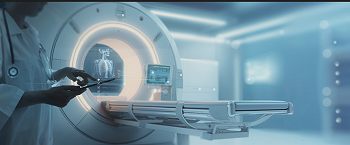
Multiparametric MRI of the prostate is a leading edge medical imaging technology. It uses a powerful magnetic field, generally 1.5 or 3.0 Tesla, and radio waves to obtain very precise images of the prostate. This examination is completely non-invasive, painless, and does not involve the use of X-rays.
The prostate MRI objectives are multiple :
- Prostate cancer screening and diagnosis: detecting the presence of suspect lesions, evaluating their precise location, measuring their size and characteristics
- Evaluation of the tumour extension: determining whether or not the tumour is localised or has progressed, checking neighbouring tissue
- Assisting treatment strategy: guiding the targeted biopsies, helping choose the most suitable treatment, defining what stage the disease has reached
- Follow-up and surveillance: checking development of a known tumour, evaluating response to treatment, detecting any return of the disease
This examination is based on a number of imaging sequences that, when combined, provide a complete analysis of the prostate. The T2 weighted sequences make it possible to observe the structure and anatomy of the prostate while the diffusion imaging helps to identify cellular abnormalities. When the injection of a contrast agent (Gadolinium) is necessary, the dynamic imaging makes it possible to study the vascularisation of tissue to better distinguish between healthy tissue and suspect tissue.
During the examination you will be asked to lie down on the imaging table and an antenna is placed on your abdomen that captures the radio signals emitted by the tissue. In rare cases an endorectal antenna may be used but this remains exceptional.
Preparation
- Check that there are no contra indications : the presence of a pacemaker, defibrillator, neurostimulators, cochlear implants, metal foreign bodies, blood sugar skin sensor, etc. Some of these devices are compatible with MRI after some preparation.
- Carry out a rectal lavage (Cleen Enema, available at chemists without prescription) either at home 1 to 3 hours before the examination or at the hospital 45 minutes before.
- In case of anxiety or claustrophobia your doctor can prescribe a sedative.
During the examination
- You will lie down on your back in a cylindrical device and must remain immobile during the examination to obtain good quality images.
- An intravenous injection of the contrast agent (Gadolinium) is often necessary.
- The MRI device makes a lot of noise that can be muffled by wearing headphones or earplugs that are supplied.
Duration
- Approximately 30 minutes
After the examination.
- The images are analysed by the radiologist who draws up a detailed report.
- An appointment with your urologist will enable you to discuss the results as a whole
The importance of expertise
A multiparametric MRI of the prostate can be carried out and the results interpreted at most radiology units in Belgium. However, a rigorous quality chain is essential to guarantee a precise diagnosis as a basis for discussing an appropriate personalised treatment.
At the Willy Grégoir Prostate Cancer Centre we apply the highest quality standards at each stage of the process as testimony to our commitment to diagnostic excellence over many years.
For optimal precision, in most cases the MRI is carried out on a device with a high magnetic field strength of 3.0 Tesla, in accordance with a standardised protocol, and with rigorous evaluation of the image quality obtained.
The image interpretation is based on international criteria (PI-RADS 2.1) for identifying suspect zones and awarding them a cancer probability score. The expertise of the radiologist who describes the MRI is crucial and criteria relating to the volume of examinations interpreted were proposed by the ESUR/ESUI consensus:
- Have interpreted at least 1 000 MRIs of the prostate.
- Interpreting at least 200 MRIs of the prostate a year.
If a suspect zone is identified our radiologists prepare the MRIs by precisely defining the prostate and suspect zone. These data are then transferred to the KOELIS Trinity® platform that is dedicated to the fusion of MRI and 3D ultrasound images, thereby guaranteeing maximum precision for the biopsy.